- Home
- Search
- Images
- Species Checklists
- US States: O-Z >
- US National Parks
- Central America
- South America
- US National Parks
- Southern Subpolar Region
|
|
|
|
Family: Physciaceae
[Rinodinella lignans Sheard ined.] |
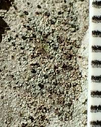
MB#484376 Type. U.S.A. COLORADO. San Juan Co., Mesa Verde National Park, Soda Canyon and Battleship Rock area, 37.14N 108.30W, on Juniperus, 2 150‑2 270 m, 29 May 1980, T.H. Nash 18126 (ASU - holotype!, SASK - isotype!). Description. Thallus crustose, thick, dark grey to dark grey-brown, areolate; areoles to 0.60-1.00 mm wide, sometimes with sublobate margins; surface plane or verrucose, shining; margin determinate or indeterminate; prothallus lacking; vegetative propagules absent. Apothecia erumpent or innate at first, becoming broadly attached, frequent, scattered or contiguous, to 0.60-0.80 mm in diam.; disc black, plane, sometimes becoming slightly convex and fissured; thalline margin concolourous with thallus or lighter at first due to broken epinecral layer, 0.05-0.15 mm wide, entire and persistent; excipular ring absent. Apothecial Anatomy. Thalline exciple 70-100 µm wide laterally; cortex 5-20 µm wide; epinecral layer 5-10 µm wide; crystals absent in cortex and medulla; cortical cells to 5.0-7.5 µm wide, pigmented; algal cells to 12.0-17.5(-30.0) µm long; cortex sometimes expanded below to 40 µm wide, cellular; proper exciple hyaline, ca. 10 µm wide laterally, expanded to (10-)20-25 µm at periphery; hypothecium hyaline, 40-80 µm deep; hymenium 85-110 µm high, not inspersed; paraphyses 2.5-3.0 µm wide, not conglutinate, with apices to 4.5-6.5 µm, darkly pigmented, immersed in dispersed pigment forming dark red-brown to brown epihymenium; asci ca. 50 x 20 µm. Ascospores 8/ascus, Type A development, Physconia-type, (15.5-)18.0-19.0(-22.0) x (8.5-)10.0-10.5(-12.0) µm, average l/b ratio 1.7-1.9, thin apical walls from start, septal thickening most evident in young spores, some spores becoming slightly constricted at maturity; torus differentiating early, becoming very darkly pigmented; walls lightly ornamented. Pycnidia immersed; conidiophores type I or VI; conidia bacilliform, 5.0-6.5 x ca. 1.5 µm. Chemistry. Spot tests all negative; secondary metabolites not detected. Substrate and Ecology. Rinodina grandilocularis has been collected on steppe shrubs, deciduous trees, conifers (most commonly Juniperus) and wood at elevations of 730-2,380 m. Substrate species include Acer grandidentatum, Artemisia cana, A. tridentata, Atriplex, Celtis, Juniperus osteosperma, Pinus cembroides and Sarcabatus verriculatus, also collected on Celtis, Pinus aristata var. longaeva and Juniperus wood. It has been collected with R. juniperina and R. lobulata. Distribution. A North American endemic species with a distribution centered on the Colorado Plateau, with outliers in Oregon and North Dakota. The locality mapped for Idaho by Sheard and Mayrhofer (2002) is an error. Notes. Rinodina grandilocularis is characterized by its Physconia-type spores with broad canals evident during development, often with uniformly thin walls (Milvina-like) at maturity. The Physcia-like stage of development is very transient and only visible as a slightly thickened apical wall in a minority of immature spores lacking wall pigmentation. Previously reported to lack a torus (Sheard and Mayrhofer 2002), a torus is differentiated around the time of first pigment deposition in the walls. It later becomes heavily pigmented and visible as a somewhat diffuse, darkly pigmented band around the septum. The species is further characterized by its grey-brown colour and typically shining surface of its well developed thallus, and by relatively large apothecia with margins lighter in colour than the thallus due to flaking of the epinecral layer. Rinodina juniperina has a similar thallus and epinecral layer, but its areoles usually possess marginal consoredia, and it has smaller, Physcia-type spores. The uniformly thin walls of mature spores lead to this species being considered a Rinodinella species early in the study and was given the herbarium name R. lignans. However, this name was never published because it became clear that the spores were too heavily pigmented to belong to the genus Rinodinella (Mayrhofer and Poelt 1978b). Specimens examined [not reported by Sheard and Mayrhofer (2002)]. U.S.A. ARIZONA. Coconino Co., Grand Canyon Nat. Park, T.H. Nash 3304, 9553b; M. Boykin 2038, 2730. COLORADO. San Juan Co., Mesa Verde Nat. Park, T.H. Nash 17898, 18125, 18126 (all ASU); Summit Co., Green Mountain Reservoir Area, I.M. Brodo 28683 (CANL). NORTH DAKOTA. Billings Co., Medora, C.M. Wetmore 45007 (MIN). OREGON. Baker Co., Lime, C. Bjorke 10595 (Goward personal herb.). UTAH. Wayne Co., Capital Reef Nat. Park, K. Yearsley C21582, C21583, C22239 (BRY). References. Sheard & Mayrhofer (2002 Fig. 12), Sheard (2004). Nash, T.H., Ryan, B.D., Gries, C., Bungartz, F., (eds.) 2004. Lichen Flora of the Greater Sonoran Desert Region. Vol 2. Thallus: crustose, thick, areolate, areoles up to 0.6-1 mm wide, sometimes with sublobate margins, plane or tumid surface: dark gray to dark gray-brown, shiny; margin: determinate or indeterminate; prothallus: lacking; vegetative propagules: absent Apothecia: erumpent or innate at first, becoming adnate, frequent, scattered or contiguous, up to 0.6-0.8 mm in diam. disc: black, plane, sometimes becoming slightly convex and fissured thalline margin: concolorous with thallus or lighter at first due to broken epinecral layer, 0.05-0.15 mm wide, entire and persistent; excipular ring: absent thalline exciple: 70-100 µm wide laterally; cortex: 5-20 µm wide; epinecral layer: 5-10 µm wide; cortical cells: up to 5-7.5 µm wide, pigmented; algal cells: up to 12-17.5(-30) µm in diam.; cortex: sometimes expanded below to 40 µm wide, cellular proper exciple: hyaline, c. 10 µm wide laterally, expanded to (10-)20-25 µm at periphery hymenium: 85-110 µm tall; paraphyses: 2.5-3 µm wide, not conglutinate, with apices up to 4.5-6.5 µm, darkly pigmented, forming dark brown epihymenium; hypothecium: hyaline, 40-80 µm thick asci: clavate, c. 50 x 20 µm, 8-spored ascospores: brown, 1-septate, broadly ellipsoid, type A development, Physconia-type, (15.5-)1819(-22) x (8.5-)10-10.5(-12) µm, thin walled from start, broad lumina canals only evident in young spores, becoming waisted at maturity; torus: absent but darkly pigmented septal wall may suggest a dark torus in mature spores; walls: ornamented Pycnidia: immersed; conidiophores: type I or VI conidia: bacilliform, 5-6.5 x c. 1.5 µm Spot tests: all negative Secondary metabolites: none detected. Substrate and ecology: on steppe shrubs, deciduous trees, conifers and wood World distribution: a North American endemic with a distribution centered on the Colorado Plateau Sonoran distribution: Arizona in Coconino and Navajo Counties at elevations of 2015-2730 m. Notes: Rinodina grandilocularis is characterized by Physconia-type spores with large lumina and thin walls from an early stage of development. The species is further characterized by the gray-brown color and typically shiny surface of its well developed thallus, and by its relatively large apothecia with margins lighter in color than the thallus due to flaking of the epinecral layer. Rinodina juniperina has a similar thallus with an epinecral layer, but its areoles usually possess marginal consoredia, and it has smaller, Physcia-type spores. |


