
- Home
- Search
- Images
- Species Checklists
- US States: O-Z >
- US National Parks
- Central America
- South America
- US National Parks
- Southern Subpolar Region
|
Acarospora malouina Øvstedal & K. Knudsen
 |
|
|
Family: Acarosporaceae
 Alan Fryday |
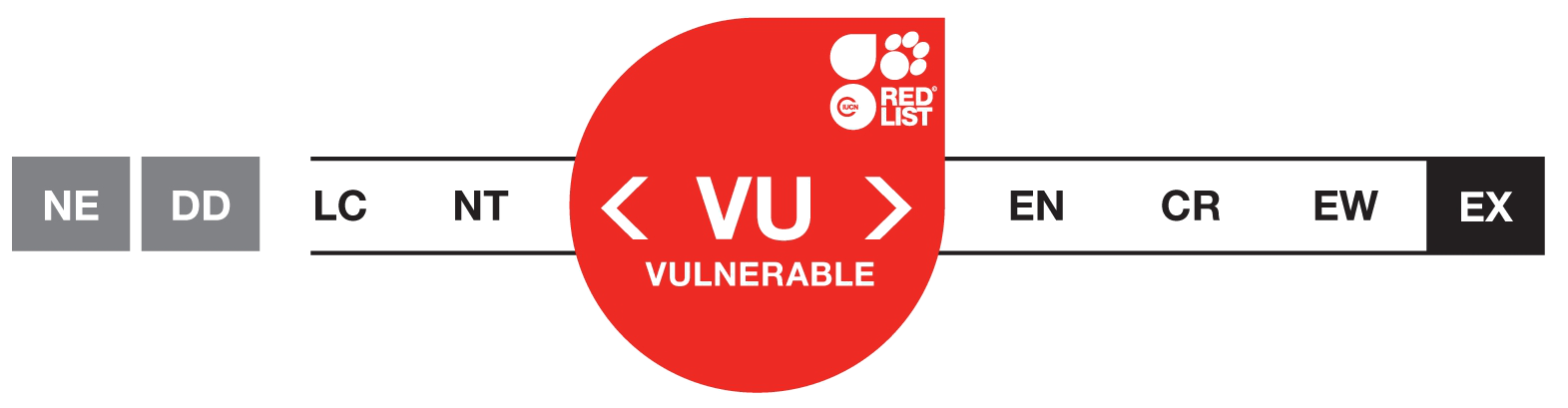
Assessed as Vulnerable D2, ver 3.1; date assessed: June 10, 2020
DOWNLOAD full IUCN Assessment as PDF Common name(s): English: n/a ASSESSMENT JUSTIFICATION [criteria: D1] Acarospora malouina is only known from two locations and its total Area of Occupancy is 8 km2. Livestock grazing and/or climatic changes could quickly lead to the decline and extirpation of this species. Therefore, it is listed as Vulnerable under criterion D2. Assessor/s: Fryday, A.; Reviewer/s: Lendemer, J.; Contributor(s): Allen, J. & Scott, T.; Facilitators(s) andCompiler(s): Allen, J. & Scott, T. Bibliography: Bachman, S., Moat, J., Hill, A.W., de la Torre, J. & Scott, B. (2011) Supporting Red List threat assessments with GeoCAT: geospatial conservation assessment tool. In: V. Smith and L. Penev (eds) e-Infrastructures for data publishing in biodiversity science. Zookeys 150: 117–126. Fryday, A. M. & Prather, L. A. (2001) The lichen collection of Henry Imshaug at the Michigan State University Herbarium (MSC). The Bryologist 104: 464-467. Fryday, A. M., Orange, A., Ahti, T., Øvstedal, D. O. & Crabtree, D. E. (2019) Checklist of lichenized and lichenicolous fungi reported from the Falkland Islands. GLALIA 8(1): 1-100. IUCN (2020) The IUCN Red List of Threatened Species. Version 2020-3. Available at: www.iucnredlist.org. (Accessed: 10 December 2020). McAdam, J. (2013) The impact of the Falklands War (1982) on the peatland ecosystem of the islands. Landscape Archaeology and Ecology 10: 143-162. Øvstedal, D. O., Lindblom, L., Knudsen, K. & Fryday, A. M. (2018) A new species of Acarospora from the Falkland Islands (Islas Malvinas). Phytotaxa 340(1): 86-92. Stenroos, S. & Ahti, T. (1992) The lichen family Cladoniaceae in the Falkland Islands. Annales Botanici Fennici 29(1): 67-73. Find out more about the IUCN Red List Categories and Criteria here. Phytotaxa 340 (1): 86–92. Thallus of contiguous squamules, 0.3–3 cm in diameter, 300–560 μm thick, indeterminate; squamules up to 2 mm wide, rugulose, fissured, continuously dividing. often forming large clusters of small squamules with 1 or 2 apothecia; upper surface glossy yellow, lower surface pale brown with a central stipe; epicortex thin, less than 10 μm thick or almost absent, of gelatinized thin hyphae, cortex red-brown throughout, 20–60 μm thick, cells usually 2–4 μm wide, angular to round; algal layer uneven, interrupted by hyphal bundles 10–60 μm wide, forming algal palisades, 150–200 μm thick, algal cells 8–12 μm wide; medulla obscure, white to brownish, 200–300 μm thick of conglutinated anticlinal hyphae mostly 1–2 μm wide, continuous with stipe. Apothecia 1–10 per squamule, disc reddish-brown, epruinose, 0.1–0.4 mm wide, with or without a distinct parathecial ring, same color as the thallus or reddish brown, darker than disc; parathecium 10–100 μm wide at surface of disc, intergrading with cortex. Layer between parathecium and hymenium I+ blue, up to two thirds of distance to disc (referred to as an abscission layer in Castello & Nimis 1994); epihymenium ca. 20–25 μm, yellowish-brown, with thin hyaline gelatinous upper layer; hymenium 200–250 μm tall, hyaline I+ blue turning red, paraphyses thin, 1–1.5(–2.0) μm wide, apices not expanded, some branching; asci 100–150 × 15–30 μm, hundreds of ascospores, ellipsoid, 2.5–3.0 × 1.5–2.0 μm; subhymenium 20–30 μm tall, I+ blue; hypothecium 20–30 μm tall, I–; pycnidia not observed, but apothecial initials abundant. Chemistry: rhizocarpic acid by tlc |
 |
|
![]()
![]()

![]()
![]()
![]()
![]()
![]()