
- Home
- Search
- Images
- Species Checklists
- US States: O-Z >
- US National Parks
- Central America
- South America
- US National Parks
- Southern Subpolar Region
|
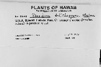
Rinodina dolichospora Malme
 |
|
|
Family: Physciaceae
 John Sheard |
MB#404311 Type. BRAZIL. MATTO GROSSO. Morro Grande, 20 December, 1893, G. Malme 2159 (lectotype, Mayrhofer et al. 1999 ‑ S!). Description. Thallus thin, ochraceous to light grey-brown, continuous to rimose-areolate, or more typically areolate; areoles discrete, spreading, to 0.30‑1.40 mm wide, often with minute lobules around the margins, lobules ca. 0.10 mm wide, sometimes becoming subsquamulose; more rarely thallus comprised of coalescing granules, 0.05-0.15 mm in diam.; surface plane, shining; margin indeterminate; prothallus lacking; vegetative propagules absent. Apothecia broadly attached to narrowly attached, frequent, rarely contiguous, to 0.60-0.90 mm in diam.; disc dark brown or black, persistently plane; thalline margin concolourous with thallus, ca. 0.10 mm wide, sometimes incomplete and/or with minute lobules similar to thallus, otherwise entire, persistent; excipular ring usually prominent, sometimes delimiting apothecium. Apothecial Anatomy. Thalline exciple 60‑90 µm wide laterally, cortex 5‑10 µm wide; epinecral layer usually present, ca. 5 µm wide; crystals absent in cortex and medulla; cortical cells to 4.0‑6.5 µm wide, pigmented or not; algal cells to 12.5‑13.5 µm long; thalline exciple 50‑70 µm wide below; proper exciple well developed, 10‑15 µm wide laterally, expanding to 30‑50 µm above, often lightly pigmented, darker when forming apothecial margin; hypothecium colourless, 60‑80 µm deep; hymenium 80‑120 µm high, not inspersed; paraphyses 2.0‑2.5 µm wide, not conglutinate, apices to 3.5‑5.0 µm wide, lightly pigmented, immersed in dispersed pigment forming a red‑brown epihymenium; asci 80‑100 x 24‑31 µm. Ascospores 8/ascus, Type A development, Pachysporaria‑type I, (18.0-) 24.5‑27.0(-34.0) x (11.0-)13.5‑15.0(-17.5) µm, average l/b ratio 1.7-2.0, many with acute apices, some lumina at first irregularly Physcia-like or subpolygonal during development, rounded at maturity, often becoming surrounded by oil small globules, sometimes with small, spherical, satellite lumina in overmature spores; torus present, narrow; walls not ornamented. Pycnidia immersed, ostioles pale brown; conidia bacilliform, 4.5-6.0 x 1.5 µm (Mayrhofer et al. 1999). Chemistry. Spot tests all negative; secondary metabolites not detected (Mayrhofer et al. 1999). Substrate and Ecology. Corticolous or lignicolous, and over mosses. Reported on Carya, Quercus alba and Q. rubra, 1,400 m in Georgia. Distribution. Rare in the southern United States, southern Mississippi valley and the west side of the Chattooga Ridge and Blue Ridge Mountains in Georgia and North Carolina, respectively. Reported from North America for the first time by Sheard & Mayrhofer (2002). The species occurs in South America and was recently recorded from Australia (Mayrhofer et al. 1999). Notes. Rinodina dolichospora is characterized by its large, Pachysporaria-type I spores, lack of secondary products and often, minute lobules of the areole margins. It is probably related to R. adirondacki by spore size and structure, the development of the proper exciple which forms a prominent excipular ring, and in the red‑brown epihymenium. It lacks pannarin, however, and has a southern distribution. Spore lumina of R. dolichospora may have an irregular polygonal shape during development and are sometimes surrounded by droplet-like inclusions in the wall at maturity (Mayrhofer et al. 1999,c). In late development the lumina become rounded and then irregularly rounded, and finally form apical satellites when overmature. Perhaps the closest relationship is with R. confinis Samp., an endemic of Portugal (Giralt 2001). This species is characterized by its granular thallus morphology, a feature only rarely developed in R. dolichospora, later developing into a subsquamulose thallus. The spores of R. confinis also possess some lumina surrounded by small oil globules but which become larger by coalescence to form satellite apical lumina in overmature spores (Giralt & Mayrhofer 1995) as described above. The two species may prove to be conspecific (Mayrhofer et al. 1999). The spreading thallus of R. dolichospora is somewhat reminiscent of R. ascociscana where however, it is more continuous. The later species is very distinct in its darker, brownish thallus, more prominent apothecial margins and its larger spores which belong to the Physcia-type. The comparison between these two species was first made by Malme (1902). Specimens examined. U.S.A. ARKANSAS. Garland Co., Hot Springs Nat. Park, C.M. Wetmore 86507 (MIN). GEORGIA. Rabun Co., Dicks Creek Gap, I.M. Brodo 23927 (CANL). LOUISIANA. Natichitoches Co., Red Dirt Wildlife Management Area, S.C. Tucker 29237a (SBBG); St. Helena Parish, 4 mi N Chipola, S.C. Tucker 18671 (LSU). NORTH CAROLINA. Jackson Co., Whiteside Mountain, T.H. Nash 18805 (ASU). Reference. Mayrhofer et al. (1999 Fig. 10). |


![]()
![]()

![]()
![]()
![]()
![]()
![]()