
- Home
- Search
- Images
- Species Checklists
- US States: O-Z >
- US National Parks
- Central America
- South America
- US National Parks
- Southern Subpolar Region
|
Rinodina innata Sheard
 |
|
|
Family: Physciaceae
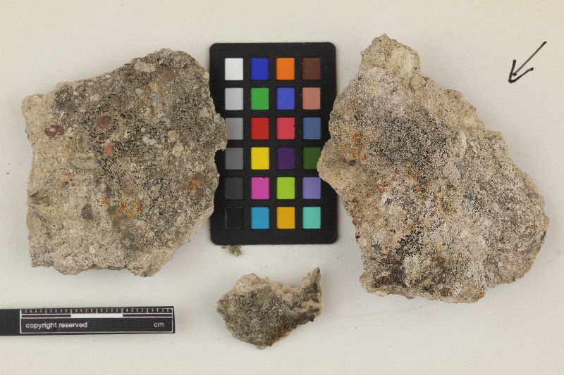 |
MB#484377 Type. U.S.A. CALIFORNIA. Santa Barbara Co., Santa Cruz Island, 7 km SSE Prisoner’s Harbor along road to radar station, 335 m, 34°00' N, 119°40'30" W, with R. gennarii, 9 January 1994, J.W. Sheard 51281a (ASU holotype!, GZU isotype!). Exsiccata. Hasse Lich. Exs. 146 (SBBG as R. confragosa). Description. Thallus crustose, grey to grey-brown, mostly thin but thicker on protected parts of substrate, areolate; areoles to 0.60-1.00 mm wide; surface plane or uneven, matt; margin indeterminate; prothallus lacking; vegetative propagules absent. Apothecia often erumpent, then innate, rarely becoming broadly or even narrowly attached, abundant, to 0.35-0.40 mm in diam., sometimes angular by compression; disc dark brown or black, sometimes concave at first, typically plane, rarely becoming slightly convex; thalline margin typically very thin, lighter than thallus due to flaking epinecral layer, <0.05-0.10 mm wide, entire and persistent; excipular ring absent. Apothecial Anatomy. Thalline exciple often not distinguishable from areole margin, 60-70 µm wide laterally; cortex ca. 5 µm wide; epinecral layer 5-15 µm wide; crystals absent in both cortex and medulla; cortical cells to 5.0-6.5 µm wide, typically lightly pigmented; algal cells to 10.0-16.5 µm long; thalline exciple ca. 80 µm below when apothecia narrowly attached; cortex not expanded; proper exciple hyaline, ca. 10 µm wide laterally, expanding to 10-30 µm at periphery; hypothecium hyaline, 50-90 µm deep; hymenium 80-100 µm high, not inspersed; paraphyses 2.0-2.5 µm wide, somewhat conglutinate, with apices to 4.0-5.0 µm, lightly to moderately brown pigmented, immersed in dispersed pigment, forming grey-, to orange-brown or reddish brown epihymenium; asci 50-60 x 16-18 µm. Ascospores 8/ascus, Type A development, Physconia-type, (13.0-)16.5-18.0 (-21.0) x (6.5-)8.5-9.5(-11.0) µm, average l/b ratio 1.8-2.0, lumina angular, frequently becoming thin walled at apices, finally losing septal wall thickening and filling cells, overmature spores often constricted; torus heavily pigmented at maturity; walls becoming heavily pigmented, not or lightly ornamented. Pycnidia rare, darkly pigmented, immersed in thallus; conidiophores type I; conidia bacilliform, 3.5-4.0 x ca. 1.0 µm. Chemistry. Spot tests all negative; secondary metabolites not detected. Substrate and Ecology. Coastal sandstone and calcareous rocks above the spray zone, sometimes on eutrophic bird perches accompanied by R. gennarii, from sea level to 1,000 m. Distribution. A North American endemic species with a Californian distribution from San Mateo Co. south to Baja California Norte. Notes. The typical form of R. innata possesses small, innate apothecia with concave to plane discs, possessing distinctively thin and light coloured margins caused by the broken epinecral layer. Specimens with thicker thalli may have an external morphology similar to R. gennarii when the apothecia become broadly attached, or rarely, narrowly attached and the discs slightly convex. Anatomically, R. innata is best distinguished by its significantly larger, Physconia-, rather than Dirinaria-type spores which develop a prominent, pigmented torus, and by the grey-, orange-, or red-brown epihymenium. Rinodina pacifica may be related to R. innata since it also possesses Physconia-type spores and both may develop endospore wall pigmentation. However, the spores of R. pacifica are significantly larger and its apothecia are always narrowly attached, and larger when mature. Rinodina innata also may be related to the Australasian R. tibellii H. Mayrhofer (Mayrhofer 1983, 1984b) which may have a similar habit but is distinguished by its larger, subinnate to broadly attached apothecia and larger, Physcia-type spores. These spores are unique within the spore type in that they inflate at the septum on application of KOH. Specimens examined. MEXICO. BAJA CALIFORNIA NORTE. Isla Cedros, 1994, T.H. Nash 34518 (ASU). U.S.A. CALIFORNIA. San Mateo Co. Menlo Park, 1966, S.C. Tucker 6344b (LSU); Santa Barbara Co. Santa Cruz Island, 1994, J.W. Sheard 5108a (SASK), Santa Cruz Island, 1994, T.H. Nash 32451 (ASU), Santa Rosa Island, 1994, J.W. Sheard 5048 (SASK), Santa Rosa Island, 1994, T.H. Nash 32835 (ASU). Nash, T.H., Ryan, B.D., Gries, C., Bungartz, F., (eds.) 2004. Lichen Flora of the Greater Sonoran Desert Region. Vol 2. Thallus: crustose, mostly thin but thicker on protected parts of substrate, areolate, areoles up to 0.6-1 mm wide, plane or uneven surface: gray to gray-brown, dull; margin: indeterminate; prothallus: lacking; vegetative propagules: absent Apothecia: often erumpent, then innate, rarely becoming adnate or even sessile, abundant, up to 0.35-0.4 mm in diam., sometimes angular by compression disc: dark brown or black, sometimes concave at first, typically plane, rarely becoming slightly convex thalline margin: typically very thin, lighter than thallus, <0.05-0.1 mm wide, entire and persistent; excipular ring: absent thalline exciple: often not distinguishable from areole margin, 60-70 µm wide laterally; cortex: c. 5 µm wide; epinecral layer: 5-15 µm wide; cortical cells: up to 5-6.5 µm wide, typically lightly pigmented; algal cells: up to 10-16.5 µm in diam.; thalline exciple: c. 80 µm below when apothecia sessile; cortex: not expanded proper exciple: hyaline, c. 10 µm wide laterally, expanding to 10-30 µm at periphery hymenium: 80-100 µm tall; paraphyses: 2-2.5 µm wide, somewhat conglutinate, with apices up to 4-5 µm, lightly to moderately brown pigmented, immersed in dispersed pigment, forming gray-, to orange-brown or reddish brown epihymenium; hypothecium: hyaline, 50-90 µm thick asci: clavate, 50-60 x 16-18 µm, 8-spored ascospores: brown, 1-septate, ellipsoid, type A development, Physcia-type, (13-)16.5-18(-21) x (6.5-)8.5-9.5(-11) µm, lumina angular, sometimes becoming rounded at ends (Physconia-like), finally filling cells when overmature, walls becoming heavily pigmented, pigmented endospore wall sometimes evident at septum, overmature spores often waisted; torus: heavily pigmented at maturity; walls: not or very lightly ornamented Pycnidia: rare, darkly pigmented, immersed in thallus; conidiophores: type I conidia: bacilliform, 3.5-4 x c. 1 µm Spot tests: all negative Secondary substances: none detected. Substrate and ecology: coastal sandstone and calcareous rocks, sometimes on eutrophic bird perches accompanied by R. gennarii above the spray zone World distribution: a North American endemic species with a southwestern oceanic distribution from central California (San Mateo Co.) south to Baja California Sonoran distribution: Channel Islands in southern California and Baja California. Notes: The typical form of R. innata possesses innate apothecia with concave to plane discs, and possesses distinctively thin and light colored margins caused by the broken epinecral layer. Specimens with thicker thalli may have an external morphology similar to R. gennarii when the apothecia become adnate, or rarely, sessile and the discs slightly convex. Anatomically, R. innata is best distinguished by its significantly larger, Physcia-, rather than Dirinaria-type spores that develop a prominent, pigmented torus and by the gray-, orange-, or red-brown epihymenium. Rinodina pacifica may be related to R. innata since it possesses Physcia-type spores in development and both develop endospore wall pigmentation. However, the spores of R. pacifica are significantly larger and its apothecia are always sessile and larger than 0.4 mm in diam. when mature. |


![]()
![]()

![]()
![]()
![]()
![]()
![]()